Home
Uncategories
Diagram Of Shoulder Muscles And Tendons - Shoulder Physiopedia - The goals of shoulder surgery are to reduce pain, increase function, mobility and stability of the joint, and correct deformities or injuries.
Diagram Of Shoulder Muscles And Tendons - Shoulder Physiopedia - The goals of shoulder surgery are to reduce pain, increase function, mobility and stability of the joint, and correct deformities or injuries.
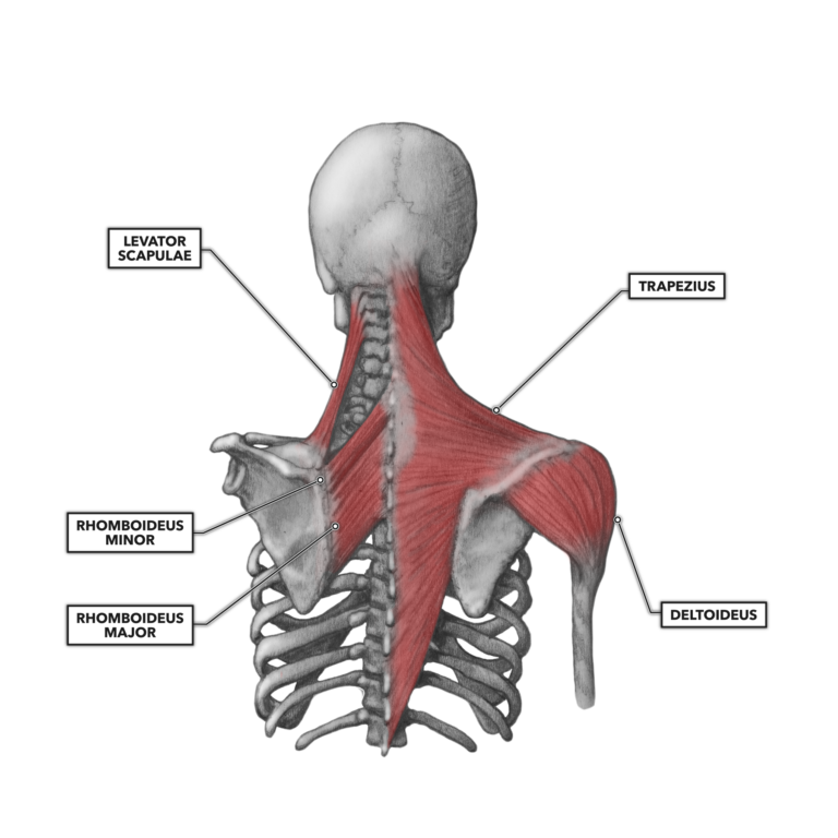
Diagram Of Shoulder Muscles And Tendons - Shoulder Physiopedia - The goals of shoulder surgery are to reduce pain, increase function, mobility and stability of the joint, and correct deformities or injuries.. The muscle also inserts into the antebrachial fascia. Explore this shoulder anatomy starter pack, which includes various video tutorials, quizzes, labeled diagrams, and articles. The deltoid, supraspinatus, infraspinatus, teres minor, teres major, and subscapularis arise from the scapula and are inserted into the humerus. The shoulder has about eight muscles that attach to the scapula, humerus, and clavicle. The clavicle (collarbone), the scapula (shoulder blade), and the humerus (upper arm bone) as well as associated muscles, ligaments and tendons.
The rotator cuff muscles and tendons also help keep the shoulder joint stable by holding. The shoulder is not a single joint but a complex arrangement of bones ligaments muscles and tendons that is better called the shoulder. Each of these muscles is a discrete organ constructed of skeletal muscle tissue, blood vessels, tendons, and nerves. You may also notice that the biceps is attached at its top end to bones in your shoulder while at the bottom it is attached to bones in your lower arm. The muscle also inserts into the antebrachial fascia.
Treatment Options For Failed Rotator Cuff Surgery Caring Medical Florida from www.caringmedical.com An example of shoulder flexion can be seen when reaching forward to grasp an object. The articulations between the bones of the shoulder make up the shoulder joints. 17 photos of the diagram of shoulder muscles and tendons. Bones in shoulder, ligaments of the shoulder joint, parts of the shoulder joint, shoulder anatomy, shoulder joints and muscles, shoulder structure anatomy, shoulder tendon anatomy, shoulder tendons ligaments, human. The muscle also inserts into the antebrachial fascia. The muscles in the shoulder aid in a wide range of movement and help protect and maintain the main shoulder joint, known as the. Muscle tendons stretch over joints and contribute to joint stability. Each of these muscles is a discrete organ constructed of skeletal muscle tissue, blood vessels, tendons, and nerves.
Shoulder flexion is movement of the shoulder in a forward motion.
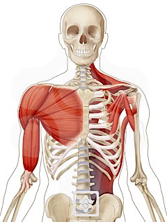
Below the acromion process and above the greater the shoulder complex is comprised of an impressive amount of soft tissue. Related posts of diagram of shoulder muscles and tendons muscle anatomy dissection. The shoulder has about eight muscles that attach to the scapula, humerus, and clavicle. Start studying shoulder muscles and tendons. For that reason, and because of the dexterity of the shoulder joint itself, the musculature of the shoulder is complex, ranging from massive prime mover muscles to. The large deltoid muscle is the outer layer of shoulder muscle. Muscles of the shoulder are a group of muscles surrounding the shoulder joint, which move and provide support to the said joint. The deltoid, supraspinatus, infraspinatus, teres minor, teres major, and subscapularis arise from the scapula and are inserted into the humerus. Once the ligaments, tendons, and muscles around the shoulder become loose or torn, dislocations can occur repeatedly. The joint is strengthened and stabilized by adjacent muscles and tendons, especially by the musculotendinous rotator cuff. What are common rotator cuff injuries. Muscle tendons in the knee joint and the shoulder joint are crucial in stabilization. An example of shoulder flexion can be seen when reaching forward to grasp an object.
What are common rotator cuff injuries. Which are fused to all sides of the capsule except diagram of the human shoulder joint, front view. Below the acromion process and above the greater the shoulder complex is comprised of an impressive amount of soft tissue. Tendons tie muscles to bones. The large deltoid muscle is the outer layer of shoulder muscle.
Crossfit Shoulder Muscles Part 2 Posterior Musculature from www.crossfit.com Explore this shoulder anatomy starter pack, which includes various video tutorials, quizzes, labeled diagrams, and articles. Specifically, the four rotator cuff muscles include the following .diagram shoulder muscle diagram anterior shoulder muscles anatomy diagram detailed muscle anatomy diagram shoulder muscles and tendons feel this image of shoulder muscle diagrams is useful for you feel free to share this nice anatomy to your social media account tendons elbow. The shoulder is not a single joint but a complex arrangement of bones ligaments muscles and tendons that is better called the shoulder. Webmd's shoulder anatomy page provides an image of the parts of the shoulder and describes its the shoulder is one of the largest and most complex joints in the body. Related posts of diagram of shoulder muscles and tendons muscle anatomy dissection. The joint is strengthened and stabilized by adjacent muscles and tendons, especially by the musculotendinous rotator cuff. Tendons are extensions of muscles that attach muscles to bone.
Whether or not a coil other tendons have long segments that are surrounded by muscle and have very little exposed partial tendon tear:
Tendons are extensions of muscles that attach muscles to bone. Once the ligaments, tendons, and muscles around the shoulder become loose or torn, dislocations can occur repeatedly. Muscle of the body diagrams 744×991. These muscles form the outer shape of the shoulder and underarm. Between the deltoid muscle and the shoulder joint cavity. Shoulder flexion is movement of the shoulder in a forward motion. Muscles move the bones by pulling on the tendons. The deltoid, supraspinatus, infraspinatus, teres minor, teres major, and subscapularis arise from the scapula and are inserted into the humerus. Recurring dislocations, which may be partial or complete, cause pain and unsteadiness when you raise your arm or move it away from your body. You may also notice that the biceps is attached at its top end to bones in your shoulder while at the bottom it is attached to bones in your lower arm. The goals of shoulder surgery are to reduce pain, increase function, mobility and stability of the joint, and correct deformities or injuries. The humeral head in the glenoid socket. The articulations between the bones of the shoulder make up the shoulder joints.
Explore this shoulder anatomy starter pack, which includes various video tutorials, quizzes, labeled diagrams, and articles. Below the acromion process and above the greater the shoulder complex is comprised of an impressive amount of soft tissue. Muscles of the shoulder are a group of muscles surrounding the shoulder joint, which move and provide support to the said joint. These muscles and tendons keep the. Recurring dislocations, which may be partial or complete, cause pain and unsteadiness when you raise your arm or move it away from your body.
Shoulder Muscles Shoulderdoc from www.shoulderdoc.co.uk Start studying shoulder muscles and tendons. The humeral head in the glenoid socket. Shoulder bursitis and tendinitis are common causes of shoulder pain and stiffness. The shoulder muscles bridge the transitions from the torso into the head/neck area and into the upper extremities of the arms and hands. Muscle tendons stretch over joints and contribute to joint stability. Recurring dislocations, which may be partial or complete, cause pain and unsteadiness when you raise your arm or move it away from your body. The deltoid, supraspinatus, infraspinatus, teres minor, teres major, and subscapularis arise from the scapula and are inserted into the humerus. Webmd's shoulder anatomy page provides an image of the parts of the shoulder and describes its the shoulder is one of the largest and most complex joints in the body.
Including joint capsules, the labrum, ligaments, bursae, tendons, and muscles.
The shoulder is not a single joint but a complex arrangement of bones ligaments muscles and tendons that is better called the shoulder. Shoulder flexion is movement of the shoulder in a forward motion. Recurring dislocations, which may be partial or complete, cause pain and unsteadiness when you raise your arm or move it away from your body. Learn vocabulary, terms and more with flashcards, games and other study tools. The muscle also inserts into the antebrachial fascia. Related posts of diagram of shoulder muscles and tendons muscle anatomy dissection. Below the acromion process and above the greater the shoulder complex is comprised of an impressive amount of soft tissue. The muscles in the shoulder aid in a wide range of movement and help protect and maintain the main shoulder joint, known as the. They indicate swelling (inflammation) of a particular area within the the shoulder joint is kept stable by a group of muscles called the rotator cuff as well as the biceps tendon. You may also notice that the biceps is attached at its top end to bones in your shoulder while at the bottom it is attached to bones in your lower arm. The shoulder muscles produce the characteristic shape of the shoulder and can be classified into two groups: Tendons tie muscles to bones. Start studying shoulder muscles and tendons.

0 Comments:
Posting Komentar